近日,Chemico-Biological Interactions(IF=5.168)在线发表了沈阳药科大学制药工程学院袁雷/生命科学与生物制药学院李艳春课题组在双重组织蛋白酶(Cathepsin)L和S抑制剂阻断胰腺癌体内外转移领域的研究成果。文章题目为“ASPER-29 suppresses the metastasis of pancreatic cancer cells by dual inhibition of cathepsin-L and cathepsin-S” (DOI: 10.1016/j.cbi.2022.109811).
肿瘤细胞转移是造成胰腺癌不良预后和高致死率的主要原因,开发有效的抗肿瘤细胞转移药物对于胰腺癌的治疗具有重要意义。课题组以研究多年的天然分子asperphenamate为先导分子,针对其分子内的苯丙氨醇片段进行结构修饰,得到化合物ASPER-29(图1),该化合物对Cathepsin L和S具有双重抑制作用,且具有较高选择性。进一步研究表明,ASPER-29在体外对两种胰腺癌瘤株PANC-1和BxPC-3显示了显著的抗转移和侵袭作用(图2)。体内研究发现ASPER-29具有明显的抗胰腺癌细胞的肝、肺转移作用(图3)。
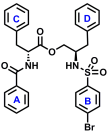
图1. The structure of ASPER-29.
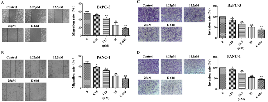
图2. ASPER-29 inhibited the migration and invasion of pancreatic cancer cells. BxPC-3 (A) and PANC-1 (B) cells were scratched by 10 μl pipet tips, and then incubated with different doses of ASPER-29 and E− 64d (25 μM) for 48 h. BxPC-3 (C) and PANC-1 (D) cells were incubated with different doses of ASPER-29 and E− 64d (25 μM), and then the cells in the upper cham- bers coated by Matrigel (0.23 mg/mL) were stained with 0.1% crystal violet to investi- gate the effect of ASPER-29 on the invasion of BxPC-3 and PANC-1 cells. The data are presented as the mean ± SD from three in- dependent experiments. *p < 0.05, **p < 0.01 vs. control group.
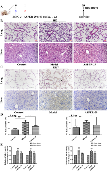
图3. ASPER-29 suppressed pancreatic cancer metastasis in vivo. (A) The construction of a tail vein metastasis model. Human BxPC-3 cells were injected into the tail vein of each mouse. One day later, ASPER-29 (100 mg/kg) was intragastrically administered once daily for eight consecutive weeks before perfusion. (B) Representative hematoxylin and eosin–stained sections of metastasis in liver and lung are shown. Original magnification: 200 × . (C) Representative IHC images displaying Ki67 expression and quantitative data in lung and liver tissues. Original magnification: 200 × . (D) Representative IHC images displaying CEACAM6 expression and quantitative data in lung and liver tissues. Original magnification: 200 × . (E) Relative CAT-L and CAT-S activities were evaluated by a BioVision’s CAT-S or CAT-L Activity Assay Kit in lung and liver tissues. The data are presented as the mean ± SD (n = 6 per group). #p < 0.05, ##p < 0.01 vs. control group; *p < 0.05, **p < 0.01 vs. model group.
本文第一作者为沈阳药科大学制药工程学院袁雷,沈阳药科大学生命科学与生物制药学院李艳春为通讯作者。该课题由辽宁省教育厅研究课题(2019LJC02)和辽宁省自然基金资助完成(20180550465)。
文章链接:https://www.sciencedirect.com/science/article/abs/pii/S0009279722000163?via%3Dihub


 location: